隐匿骨折(OF)指常规X线检查不能发现或疑似但不能明确诊断,需其他特殊检查才能发现的骨折[1]。强直性脊柱炎(AS)患者脊椎强直后力学特性发生变化,轻微暴力就可能导致其骨折[2-3]。本院曾收治2例AS并颈椎隐匿不全骨折患者,确诊后给予非手术治疗,未进展到完全骨折。现将诊疗过程报告如下。
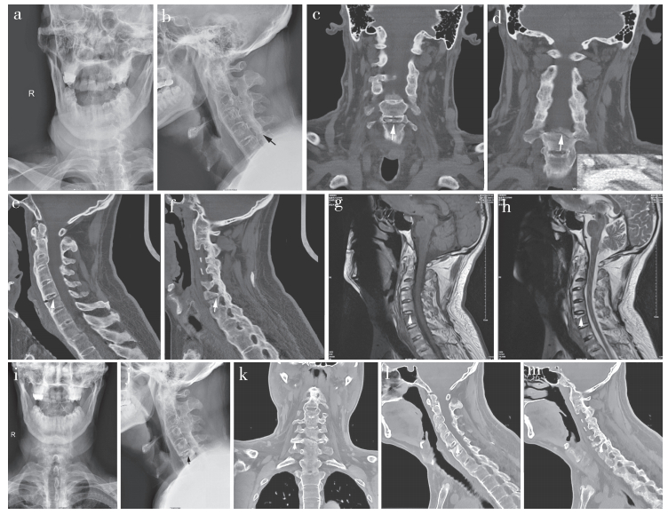
1 临床资料 1.1 病例1颈型AS患者,男,52岁。头颈部活动受限30余年。因足部被障碍物绊到,头部碰撞前方物体后出现颈痛来本院就诊。查体:面容痛苦,颈部不可屈伸及旋转。自右侧颈动脉鞘与内脏鞘间隙按压椎体前方,在喉结下方2 cm处有明显压痛,颈椎纵向叩击痛阳性,与压痛点部位重合,神经功能正常。X线片示颈椎竹节样变,生理曲度变直,重心前移,C5,6关节突关节区域似可见透亮线(图 1a,b)。疑C6关节突骨折,因患者个人原因未作进一步检查。给予止痛药及颈托围领制动。建议2~3 d后疼痛无缓解复诊。

|
a,b:颈椎X线片示AS,未见椎前软组织阴影增宽,侧位X线片C5,6关节突关节区域似可见透亮线 c~f:颈椎CT(骨窗)提示C5/C6椎间盘有连续性骨痂通过,C6上终板前方大部同椎体分离(c,e),但关节突关节已经骨性融合(d),右侧椎弓根亦有骨折线通过(f) g,h:颈椎MRI示C6椎体上部T1WI为低信号,T2WI为高信号,提示新鲜损伤,其他区域骨质及软组织信号正常,提示不完全性C6椎体骨折,无其他颈椎区域软组织及骨结构损伤 i,j:治疗后3个月复查X线片,C6上终板下方可认出骨折线,提示骨折周围骨形成和吸收同时发生k~m:治疗后3个月复查CT,可见骨折线裂隙增大同时连续性骨痂通过,提示骨形成与骨吸收同时发生 图 1 典型病例影像学资料 |
患者口服止痛药及佩戴颈托后颈痛略减轻。3 d后因乘车时颈前部疼痛加重而来院复诊。查体同前。行颈椎CT扫描,显示C6上终板与椎体分离,骨折线延伸至右侧椎弓根(图 1c~f);诊断为C6经椎体不全骨折[4]。
MRI示C6椎体前皮质在C6上终板下方连续性中断,终板下方骨质T1WI呈低信号,T2WI为高信号(图 1g,h)。明确C6为新鲜不全骨折。继续给予颈托围领制动,告诫患者治疗期间避免乘车,避免颈部遭受外力。
1.2 病例2AS患者,男,75岁。10年前曾因胸椎骨折行内固定手术。因乘车被追尾后颈痛就诊。查体:表情痛苦,颈项部各项活动消失,自右侧颈动脉鞘与内脏鞘间隙向后方按压颈椎体前方,在喉结下方4 cm处有明显压痛,颈椎纵向叩击痛阳性,与压痛点一致。X线片示颈椎竹节样变,曲度变直。冠状位CT示颈椎略向右侧凸,C7上终板与椎体间形成一横贯并纵贯椎体全长的骨折线,前缘略张开,后方的关节突关节呈融合状态。MRI示C7上终板下新鲜骨折,T1WI为低信号,T2WI呈高信号,脊髓信号正常。后方骨及软组织结构正常。明确为C7经椎体型新鲜不全骨折。给予佩戴颈托围领制动。定期随访。
2 结果病例1患者伤后2周颈痛消失,但有轻度按压痛点及纵向叩击痛。伤后6周,压痛及纵向叩击痛消失,但去除颈托并尝试颈椎过伸动作时有轻度不适。继续佩戴颈托2周。术后3个月复查X线片,C6上终板下可辨出骨折线(图 1i,j)。CT示骨折线较前增宽,无移位及成角,部分区域有连续性骨痂通过(图 1k~m)。病例2伤后3周复诊,颈部压痛及纵向叩击痛均消失,建议继续佩戴颈托5周。均继续随访中,告诫患者避免外伤。
3 讨论 3.1 AS患者轻微暴力可致脊椎骨折正常脊柱关节能吸收多方向应力而使脊椎受到最小伤害。AS患者后期脊柱僵硬强直、力臂延长、活动度丧失[2-3];骨质脆性增加及骨质疏松(骨量减少者19%~62%,骨质疏松者13%~16%)[5-6];颈椎受累后视觉范围受限,前庭及本体感觉障碍,易摔倒[7];微小暴力就可致骨折[8];脊椎骨折有56.0%~58.6%由无/轻至中度创伤引起,47.0%~78.2%伴脊髓损伤[9]。Prieto-Alhambra等[10]报道AS患者骨折比例达0.11%(139/124 655),同期正常人群为0.07%(271/373 962),其中脊椎骨折的风险比同期正常人群高5倍。Robinson等[11]报道瑞典国立医院12年间接收17 764例AS患者,脊柱骨折率为4.1%。由于肌肉保护弱及椎体周围软组织骨化,下颈椎为骨折的好发部位[12]。Vosse等[13]报道AS患者中约0.4%伴脊椎骨折,其中颈椎骨折占36.0%。Lukasiewicz等[2]报道颈椎骨折占脊椎骨折的53.0%。颈椎骨折以C6,7最多,C5,6其次[14],多为过伸损伤引起的三柱不稳骨折,脊髓受损占21.0%~92.0%[2-3, 5, 8]。
3.2 如何发现无脊髓损伤的AS颈椎隐匿骨折AS颈椎骨折患者早期大多神经障碍轻微,不易联想到骨折,而常规影像辨别效果差,故易漏诊[15]。解剖标志变化、韧带骨化及假象等是常规影像漏诊的重要原因[16]。Gilard等[3]报道7例,3例漏诊。苏格兰伊丽莎白国立脊柱损伤中心12年收治32例AS颈椎骨折患者,15例伤后无脊髓损伤表现,19例(59.4%)常规影像漏诊[16]。
根据临床经验及文献总结可从3个方面判断AS患者是否发生颈椎骨折:①详细了解损伤机制,颈椎骨折多为过伸损伤,一旦有该机制的损伤即使暴力微小,也要考虑骨折可能[3, 5, 8]。②症状可仅表现为颈痛[16-17]。③临床查体痛点明确,且压痛较剧。前柱骨折时颈椎前方压痛为主,尝试仰头时疼痛加重;波及后柱时项部压痛较剧,尝试低头时疼痛加重。但后柱骨折部位深在,压痛阴性时也不能排除。纵向叩击痛多阳性,与压痛部位一致。如软组织损伤,疼痛多较轻,纵向叩击痛多阴性。一旦怀疑骨折,需即刻制动保护,并行CT及MRI检查[1, 3]。
AS颈椎隐匿骨折分不完全性和完全性。前者未波及三柱,属稳定性,如处置不当,可进展为完全骨折[18];后者波及三柱,不稳定,可继发移位及脊髓损伤[3, 16]。二者鉴别:前者疼痛较为局限,后者较广泛。后者颈部尝试屈伸动作时均有疼痛加重,有时颈部有错动感,可有一过性沿神经根的放射痛;纵向叩击时颈前部及项部均有疼痛。查体要轻柔,结果仅供参考。一旦怀疑,均应先按不稳定骨折处理。
CT及MRI检查各有所长。CT可诊断骨性损伤,明确是否为完全骨折;MRI能发现血肿、脊髓损伤、软组织损伤、骨挫伤及辨别是否新鲜骨折,且可评判骨折稳定性[1]。本组2例病例CT明确为不全骨折,MRI明确骨折为新发,属稳定性,为临床制定治疗方案提供了客观依据。CT或MRI也可呈假阴性。Koivikko等[19]报道MRI发现2例CT漏诊的颈椎骨折,而多层螺旋CT发现6例MRI漏诊的颈椎骨折。
健康人群无移位颈椎骨折,可有椎前软组织阴影增宽[20]。本组病例的AS椎前软组织阴影正常。分析原因:①椎旁软组织钙化,肿胀基础大为削弱[21];②微小暴力即骨折,软组织尚未达到损伤临界值;③多为过伸损伤,非直接暴力,软组织损伤轻。
3.3 AS颈椎骨折的分型Glace等[4]将脊椎骨折分为经椎间盘型和经椎体型。Koivikko等[19]报道20例颈椎骨折,经椎间盘型7例。Caron等[22]将脊椎骨折分为4型:A型为经椎间盘型;B型为经椎体型;C型为前部通过椎体;后部通过椎间隙;D型与C型相反。Ma等[14]据此将AS颈椎骨折归为Ⅲ型,Ⅰ和Ⅱ型分别为无移位的经椎间盘型和经椎体型,Ⅲ型为经椎间盘或经椎体,有明显移位;分别占12例,5例和8例。之所以有经椎间盘型,是由于椎间盘钙化形成竹节样连接。笔者认为,AS早期椎间盘钙化强度不足,而椎体骨质疏松尚不严重,骨折以椎间盘型多见。AS成熟期,大量连续性骨痂通过椎间盘,钩椎关节融合后更似骨皮质结构,而椎体多有骨质疏松,致椎间连接强度大于椎体本身,故经椎体骨折比例上升。本组2例均为AS成熟期,故骨折均为经椎体型。
3.4 无脊髓功能障碍的AS颈椎隐匿不全骨折的治疗选择AS颈椎骨折并脊髓损伤需手术治疗[1],非手术治疗限于骨折稳定、三柱未全受累且序列正常者[23]。无脊髓受损的不稳定骨折,多认为需手术。但AS患者多有骨质疏松,手术并发症多,也有学者持慎重意见[5]。Graham等[8]对AS脊柱不稳定骨折行非手术治疗,均骨性融合,并发症发生率及死亡率明显低于手术组。Mathews等[24]报道11例AS脊柱骨折,6例手术治疗,3例脊髓障碍加重(2例术前已发生);非手术治疗5例,3例神经功能维持正常,1例完全恢复,1例恶化。但不稳定骨折可发生再移位、假关节形成、迟发血肿及脊髓功能障碍等,需慎重[25],颈托围领制动也有骨折移位风险[26]。
本组2例患者选择非手术治疗。原因:①为稳定型不全骨折。②无脊髓受压。③经椎体型骨折,骨质疏松明显,钛板固定可能发生螺钉松动等并发症;术中麻醉及搬动患者,可能进展为医源性完全骨折,甚至脊髓损伤[2-3]。④AS患者韧带骨赘、骨膜及椎骨多同时存在自我新骨形成,有助骨折愈合[27]。有报道非手术治疗骨性融合需5个月[28]。⑤无颈椎后凸,颈托围领不会加重骨折[21]。
本组2例颈托围领制动3~6周后,局部症状消失,随访期无新发症状。但病例1伤后3个月X线片可清晰看到骨折线,应为AS患者骨吸收与骨形成同时发生所致[29]。说明X线片在随访复查非常有价值:可明确骨折线是否有吸收;便于早期发现是否进展为完全骨折并出现不稳及移位,避免单靠症状及体征判断而可能出现的漏诊[25]。AS患者,轻微外力可致颈椎骨折,可以推测,在已有骨折时,小于原有外力阈值的暴力可能导致不全骨折进展为完全骨折甚至移位,后者X线片容易发现。
CT可明确是否骨性愈合[30]。病例1术后3个月CT示骨折间隙增大,但骨折线内有连续性骨痂通过,为骨形成与骨吸收同时发生的表现。骨折线未波及全层,仍属稳定骨折。患者无新发疼痛,提示纤维骨痂初步维持了骨折稳定,但强度不足,应继续采取保护措施。患者仍需密切随访,一旦进展为完全骨折或脊髓受压,仍需手术减压和重建脊柱稳定性。
| [1] | Elgafy H, Bransford RJ, Chapman JR. Epidural hematoma associated with occult fracture in ankylosing spondylitis patient:a case report and review of the literature[J]. J Spinal Disord Tech, 2011, 24(7): 469–473. DOI:10.1097/BSD.0b013e318204da02 |
| [2] | Lukasiewicz AM, Bohl DD, Varthi AG, et al. Spinal fracture in patients with ankylosing spondylitis:cohort definition, distribution of injuries, and hospital outcomes[J]. Spine (Phila Pa 1976), 2016, 41(3): 191–196. DOI:10.1097/BRS.0000000000001190 |
| [3] | Gilard V, Curey S, Derrey S, et al. Cervical spine fractures in patients with ankylosing spondylitis:Importance of early management[J]. Neurochirurgie, 2014, 60(5): 239–243. DOI:10.1016/j.neuchi.2014.06.008 |
| [4] | Glace B, Dubost JJ, Ristori JM, et al. Transversal fractures in spinal ankylosis:a case series of 17 patients[Article in French][J]. Rev Med Interne, 2011, 32(5): 283–286. DOI:10.1016/j.revmed.2010.10.356 |
| [5] | El Tecle NE, Abode-Iyamah KO, Hitchon PW, et al. Management of spinal fractures in patients with ankylosing spondylitis[J]. Clin Neurol Neurosurg, 2015, 139: 177–182. DOI:10.1016/j.clineuro.2015.10.014 |
| [6] | van der Weijden MA, Claushuis TA, Nazari T, et al. High prevalence of low bone mineral density in patients within 10 years of onset of ankylosing spondylitis:a systematic review[J]. Clin Rheumatol, 2012, 31(11): 1529–1535. DOI:10.1007/s10067-012-2018-0 |
| [7] | Fatemi G, Gensler LS, Learch TJ, et al. Spine fractures in ankylosing spondylitis:a case report and review of imaging as well as predisposing factors to falls and fractures[J]. Semin Arthritis Rheum, 2014, 44(1): 20–24. DOI:10.1016/j.semarthrit.2014.03.007 |
| [8] | Graham B, Van Peteghem PK. Fractures of the spine in ankylosing spondylitis. Diagnosis, treatment, and complications[J]. Spine (Phila Pa 1976), 1989, 14(8): 803–807. DOI:10.1097/00007632-198908000-00005 |
| [9] | Mahajan R, Chhabra HS, Srivastava A, et al. Retrospective analysis of spinal trauma in patients with ankylosing spondylitis:a descriptive study in Indian population[J]. Spinal Cord, 2015, 53(5): 353–357. DOI:10.1038/sc.2014.150 |
| [10] | Prieto-Alhambra D, Muñoz-Ortego J, De Vries F, et al. Ankylosing spondylitis confers substantially increased risk of clinical spine fractures:a nationwide case-control study[J]. Osteoporos Int, 2015, 26(1): 85–91. DOI:10.1007/s00198-014-2939-3 |
| [11] | Robinson Y, Sandén B, Olerud C. Increased occurrence of spinal fractures related to ankylosing spondylitis:a prospective 22-year cohort study in 17764 patients from a national registry in Sweden[J]. Patient Saf Surg, 2013, 7(1): 2. DOI:10.1186/1754-9493-7-2 |
| [12] | Jo DJ, Kim SM, Kim KT, et al. Surgical experience of neglected lower cervical spine fracture in patient with ankylosing spondylitis[J]. J Korean Neurosurg Soc, 2010, 48(1): 66–69. DOI:10.3340/jkns.2010.48.1.66 |
| [13] | Vosse D, Feldtkeller E, Erlendsson J, et al. Clinical vertebral fractures in patients with ankylosing spondylitis[J]. J Rheumatol, 2004, 31(10): 1981–1985. |
| [14] | Ma J, Wang C, Zhou X, et al. Surgical therapy of cervical spine fracture in patients with ankylosing spondylitis[J]. Medicine (Baltimore), 2015, 94(44): e1663. DOI:10.1097/MD.0000000000001663 |
| [15] | Thumbikat P, Hariharan RP, Ravichandran G, et al. Spinal cord injury in patients with ankylosing spondylitis:a 10-year review[J]. Spine (Phila Pa 1976), 2007, 32(26): 2989–2995. DOI:10.1097/BRS.0b013e31815cddfc |
| [16] | Anwar F, Al-Khayer A, Joseph G, et al. Delayed presentation and diagnosis of cervical spine injuries in long-standing ankylosing spondylitis[J]. Eur Spine J, 2011, 20(3): 403–407. DOI:10.1007/s00586-010-1628-y |
| [17] | Ambrose NL, Cunnane G. Importance of full evaluation in patients who complain of neck pain[J]. Ir J Med Sci, 2009, 178(2): 209–210. DOI:10.1007/s11845-008-0256-6 |
| [18] | Backhaus M, Citak M, Kälicke T, et al. Spine fractures in patients with ankylosing spondylitis:an analysis of 129 fractures after surgical treatmen[t Article in German][J]. Orthopade, 2011, 40(10): 917–924. DOI:10.1007/s00132-011-1792-8 |
| [19] | Koivikko MP, Koskinen SK. MRI of cervical spine in juries complicating ankylosing spondylitis[J]. Skeletal Radiol, 2008, 37(9): 813–819. DOI:10.1007/s00256-008-0484-x |
| [20] | Moch AL, Schweitzer ME, Parker L. Prevertebral soft tissue swelling following trauma:usefulness following tube placement[J]. Skeletal Radiol, 2000, 29(6): 340–345. DOI:10.1007/s002560000197 |
| [21] | Clarke A, James S, Ahuja S. Ankylosing spondylitis:inadvertent application of a rigid collar after cervical fracture, leading to neurological complications and death[J]. Acta Orthop Belg, 2010, 76(3): 413–415. |
| [22] | Caron T, Bransford R, Nguyen Q, et al. Spine fractures in patients with ankylosing spinal disorders[J]. Spine (Phila Pa 1976), 2010, 35(11): E458–464. DOI:10.1097/BRS.0b013e3181cc764f |
| [23] | An SB, Kim KN, Chin DK, et al. Surgical outcomes after traumatic vertebral fractures in patients with ankylosing spondylitis[J]. J Korean Neurosurg Soc, 2014, 56(2): 108–113. DOI:10.3340/jkns.2014.56.2.108 |
| [24] | Mathews M, Bolesta MJ. Treatment of spinal fractures in ankylosing spondylitis[J]. Orthopedics, 2013, 36(9): e1203–1208. DOI:10.3928/01477447-20130821-25 |
| [25] | Aoki Y, Yamagata M, Ikeda Y, et al. Failure of conservative treatment for thoracic spine fracture in ankylosing spondylitis:delayed neurological deficit due to spinal epidural hematoma[J]. Mod Rheumatol, 2013, 23(5): 1008–1012. DOI:10.3109/s10165-012-0726-6 |
| [26] | Nahed BV, Walcott BP, Ortman AJ, et al. Interval, acute onset airway obstruction associated with a fracture of the C4 vertebra in a patient with ankylosing spondylitis[J]. J Clin Neurosci, 2010, 17(8): 1085–1088. DOI:10.1016/j.jocn.2009.12.010 |
| [27] | Sambrook PN, Geusens P. The epidemiology of osteoporosis and fractures in ankylosing spondylitis[J]. Ther Adv Musculoskelet Dis, 2012, 4(4): 287–292. DOI:10.1177/1759720X12441276 |
| [28] | 康意军, 陈飞, 吕国华, 等. 强直性脊柱炎颈椎骨折的治疗[J]. 中国脊柱脊髓杂志, 2006, 16(6): 424–428. |
| [29] | Mayle RE Jr, Cheng I, Carragee EJ. Thoracolumbar fracture dislocation sustained during childbirth in a patient with ankylosing spondylitis[J]. Spine J, 2012, 12(11): e5–8. DOI:10.1016/j.spinee.2012.10.012 |
| [30] | Nakstad PH, Server A, Josefsen R. Traumatic cervical injuries in ankylosing spondylitis[J]. Acta Radiol, 2004, 45(2): 222–226. DOI:10.1080/02841850410004085 |
 2017, Vol.15
  Issue(1): 57-60, 64


