2. 福建医科大学附属龙岩第一医院手术室, 龙岩 364000
2. Department of Operating Room, Longyan First Hospital Affiliated to Fujian Medical University, Longyan 364000, Fujian, China
在椎弓根螺钉内固定术中, 椎弓根螺钉内偏进入椎管可能引起急性或迟发性脊髓神经损伤等严重并发症[1]。为了及时发现问题并予以纠正, 术者往往会在置钉后或术后通过X线片来评估钉道情况, 目前通用的评价方法(传统方法)是以正位X线片上椎弓根螺钉尖端不超过棘突中线为内偏正常标准, 此方法主观性较大, 评定结果不仅与螺钉本身方向有关, 同时还受螺钉长度、射线投照角度及棘突解剖情况等影响[2]。本研究设计了一种采用正侧位X线片判断椎弓根螺钉内偏情况的新方法, 并通过与传统方法比较评价其诊断价值, 现报告如下。
1 资料与方法 1.1 一般资料选取2011年1月—2017年3月行椎弓根螺钉内固定术治疗的患者175例, 其中男78例、女97例, 年龄14 ~ 83(44.9±29.8)岁; 脊柱骨折49例、脊柱退行性疾病67例、脊柱肿瘤21例、脊柱感染12例、脊柱畸形26例。入选病例共置入1 099枚螺钉, 术后均接受正侧位X线及CT复查。图像入选标准:①X线图像清晰, 能清楚显示侧位椎体后缘及正位椎弓根内缘等标志; ②正位X线片上椎弓根螺钉方向朝向棘突, 且投影长度 > 1 cm; ③CT图像能准确判断椎弓根螺钉内偏情况。符合入选标准的椎弓根螺钉影像共844枚(76.8%)。置钉节段为T3 ~ S1, 其中T3 4枚, T4 12枚, T5 22枚, T6 16枚, T7 28枚, T8 19枚, T9 36枚, T10 61枚, T11 56枚, T12 99枚, L1 39枚, L2 62枚, L3 84枚, L4 114枚, L5 131枚, S1 61枚。
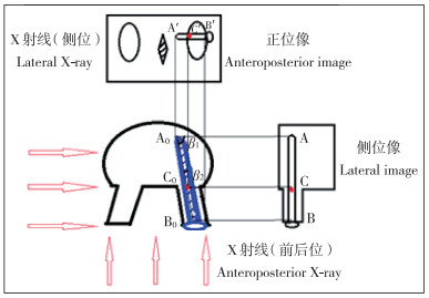
1.2 新测量方法螺钉不过度内偏穿入椎管的X线影像判断法:当侧位X线片上螺钉进入椎体后缘时, 正位X线片上螺钉尖端不能超过椎弓根内缘[3-5]。但术后正位X线片上螺钉前端已超出椎弓根影并接近椎体中央, 无法直观体现螺钉尖端刚到达椎弓根前端(椎体后缘)时的投影点(目标投影点)。为此, 本研究通过图像等比投像原理来推算目标投影点。如图 1所示, 螺钉尖端A0在正侧位X线片上投影点分别为A′和A, 螺钉末端B0点在正侧位X线片上投影点分别为B′和B; 螺钉中心轴与椎体后缘交界点为C0点, 则其侧位X线片上投影点C正好位于螺钉影与椎体后缘影的交点, 而所需推算的正位X线片上的目标投影点C′为C0的正位X线片投影点。螺钉在正侧位X线片上的投影长度受螺钉实际长度、投照角度β(设定侧位射线与螺钉方向夹角为β1, 正位为β2)及投照放大倍率n(n与球管投照距离及屏幕放大倍数等有关, 设定侧位投照放大倍率为n1, 正位为n2)等3个因素的影响。螺钉投影长度公式:侧位X线片上AC=A0C0×Sinβ1×n1, AB=A0B0×Sinβ1×n1, 故AC/AB=A0C0/A0B0; 正位X线片上A′C′=A0C0×Sinβ2×n2, A′B′=A0B0×Sinβ2×n2, 故A′C′/A′B′=A0C0/A0B0, 依等比关系可得AC/AB=A′C′/A′B′。因此, 要想在X线片上标记出目标点C′, 则需要计算出A′C′长度。这在操作上仅需3个步骤:①在侧位X线片上, 以螺钉尖端起点为A, 设定连接杆与螺钉中心轴交界处为螺钉末点B, 测量AB距离(图 2a); 设定螺钉中心轴与椎体后缘交界处为C, 则AC代表螺钉在椎体内的长度(图 2b), 计算AC与AB比值r。②在正位X线片上, 以螺钉起点为A′, 设定螺钉中心轴与连接杆中心轴连接的交点为螺钉末点B′(图 2c), 测量A′B′长度, 乘以r后得到A′C′长度, 则C′点即为目标投影点(图 2d)。③C′点与椎弓根影内侧壁距离用d值表示。C′点位于椎弓根影内侧缘的外侧, 则d值为负, 判定螺钉不内偏; 如位于椎弓根影内侧缘的内侧, 则d值为正, 判定螺钉内偏。

|
注:设定螺钉尖端为A0点、螺钉头端为B0点、螺钉中心轴与椎体后缘交界点为C0点; 在侧位X线片上, A0点投影点为A点、B0点投影点为B点、C0点投影点为C点; 测量AB及AC距离, 计算AC与AB比值r; 在正位X线片上, A0投影点为A′点、B0点投影点为B′点, 测量A′B′长度, 以A′B′×r的值作为A′C′长度, C′点为目标投影点 Note:Screw tip is set as point A0, screw head as point B0, and junction of screw's center shaft and vertebral posterior margin as point C0; On lateral roentgenograph, projection point of A0 is A, and projection point of B0 is B, and projection point of C0 is C; Distance of AB and AC as well as the specific value r(AC/AB) are calculated; On anteroposterior roentgenograph, projection point of A0 is A′, and projection point of B0 is B′; Through measuring distance of A′B′ and then multiplying ratio r, distance of A′C′ is acquired, then point C′ is target projection point 图 1 新方法设计原理 Figure 1 Design principle of new method |

|
a:AB=42.45 mm b:AC=20.20 mm, r=0.476 c:A′B′=22.34 mm d:A′C′=22.34 mm×0.476=10.63 mm; C′点位于椎弓根内壁内侧, 判断为螺钉内偏e:术后CT证实螺钉内偏 a:AB=42.45 mm b:AC=20.20 mm, r=0.476 c:A′B′=22.34 mm d:A′C′=22.34 mm×0.476=10.63 mm; target projection point C′ locating inside medial margin of vertebral pedicle, and internal shifting of pedicle screws is judged as abnormal e:Postoperative CT verifies internal shifting of pedicle screws is abnormal 图 2 新方法测量椎弓根螺钉内偏情况 Figure 2 New method to judge internal shifting of pedicle screws |
由3名不同年资的脊柱外科医师(分别具有12年、6年及4年专科工作经验)分别独立对844个螺钉的图像进行判断, 每个螺钉的图像均采用2种方法进行评估。为了减少主观偏倚, 要求对同1个螺钉图像的2种方法判断时间间隔 > 3 d。结果按内偏情况判断为是或否; 将2名或3名医师判断一致的结果作为认定结果。图像分析及测量均在福建医科大学附属龙岩第一医院PACS图像平台上进行。由3名高年资的放射科医师(分别具有12年、13年、28年影像诊断工作经验)利用术后横断面CT图像对上述844个螺钉进行判断(图 2e), 将2名或3名医师判断一致的结果作为金标准。按Aoude等[6]推荐的螺钉内偏分级标准记录结果:0级, 螺钉完全在椎弓根内; Ⅰ级, 螺钉突破椎管内侧壁最大距离 < 2 mm; Ⅱ级, 螺钉突破椎管内侧壁最大距离为≥2 mm且≤4 mm; Ⅲ级, 螺钉突破椎管内侧壁最大距离 > 4 mm。
1.4 统计学处理应用SPSS 19.0软件对数据进行统计学分析, 计数资料及率的比较采用χ2检验; 以P < 0.05为差异有统计学意义。
2 结果3名脊柱外科医师均成功完成了X线片测量。3名医师使用新方法判断螺钉内偏情况的观察者间一致率为90.5%(764/844), 而传统方法为71.2%(601/844), 差异有统计学意义(P < 0.05)。根据CT图像进行评估, 共91枚内偏螺钉, 753枚非内偏螺钉。新方法及传统方法在753枚非内偏螺钉中检测结果相符的数量分别为679枚及587枚, 在91枚内偏螺钉中检测结果相符的数量分别为86枚及61枚, 新方法在诊断灵敏度、特异度、约登指数、阳性似然比、阴性似然比等指标上均优于传统方法, 差异有统计学意义(P < 0.05, 表 1)。新方法在以CT为依据评估的0、Ⅰ、Ⅱ及Ⅲ级螺钉内偏中检测准确率分别为90.2%、92.3%、94.7%、100%, 均优于传统方法(分别为78.0%、59.0%、73.7%、71.4%), 差异有统计学意义(P < 0.05, 表 2)。
|
|
表 1 2种方法的诊断效能 Table 1 Clinical diagnostic value of 2 methods |
|
|
表 2 2种方法的准确性 Table 2 Accuracy of 2 methods |
Aoude等[7]调查发现, 目前仍有近75%的脊柱外科医师在术中使用X线透视来检测并调整螺钉位置, 但术后CT检查时依然会发现置钉存在一定的失误率, 而其中失误率最高也最让医师担忧的是螺钉过于内偏。目前临床上较通用的判断方法(传统方法)是正位X线片上螺钉尖端不超过棘突中线为内偏正常。刘浩等[2]依据此法来判断螺钉位置, 发现经验不足的医师判断错误率达23.9%, 而经验丰富者亦达17.7%;术中检出定位正确螺钉的可靠性高, 而检出错误螺钉的可靠性低。螺钉尖端投影结果与诸多因素有关。①入钉点位置及水平面倾角:入钉点越靠内、内倾角越大, 投影点越偏内。②螺钉长度:螺钉越长其在椎体内距离也越长, 投影点也越接近棘突。此时, 对于不内偏的螺钉, 可能由于螺钉过长, 投影点越过中线, 被误判为内偏螺钉; 反之, 对于内偏螺钉, 可能由于螺钉较短, 投影点仍位于同半侧区域, 而被误判为正常螺钉。③射线投照角度:当投照源往一侧偏斜时, 同侧螺钉投影长度变短, 对侧变长, 两者与棘突间的距离也发生了相应改变[8]。此外, 对于棘突偏斜畸形或因术中减压致棘突缺如等病例, 此判断方法可能会造成更多的误差。
正确置钉要求螺钉始终在椎弓根内前进。有学者提出螺钉不过度内偏穿入椎管的X线影像判断法:当侧位X线片上螺钉进入到椎体后缘时正位X线片上螺钉尖端不能超过椎弓根内缘[3-5]。这一判断方法在影像上客观、诊断价值高、可重复性好。本研究的新方法正是借助了此种判断依据。然而, 椎弓根螺钉置入后正位X线片上螺钉前端已超出椎弓根影并接近椎体中央, 无法直观体现螺钉尖端刚到达椎弓根前端(椎体后缘)时的投影点(目标投影点)。为此, 本研究通过图像等比投像原理来推算目标投影点, 根据目标投影点与椎弓根影内缘的位置关系判断螺钉是否内偏。
新方法操作简单快速, 无需特殊设备, 测量结果的准确性有赖于精确找到目标投影点(C′点)。测量时需要注意如下细节。①正侧位X线片上螺钉末点(即B点及B′点)的确定。实验设定末点为连接杆与螺钉中心轴的交点, 将此标记点在实物螺钉上标记后通过反复正侧位透视观察得出了本实验的B点及B′点定义。如能在螺钉末端设计一个可在影像上显示的标记点则能进一步提高2点的吻合性。②侧位X线片上椎体后缘线的确定。如果侧位X线片投照角度标准, 则后缘为一条垂线, 容易确定; 但当投照不标准时, 则后缘可能呈现为2条垂线, 此时需结合螺钉、连接杆投照的形态来判断2条垂线分别代表哪侧的后缘, 避免发生测量错误。③图像处理技术。利用PACS系统工具对图像进行亮度及对比度调整, 以便清晰显示椎体后缘、椎弓根影像及螺钉交界面影像等; 同时通过放大镜工具对图像进行放大后测量, 尽可能提高测量精度。
由于侧位X线片上T1和T2甚至部分T3及T4椎体锁骨及肩关节阻挡, 其椎体后缘无法清晰显示, 本实验主要纳入T3 ~ S1节段图像。实验发现理想的螺钉C′点主要分布在椎弓根影内缘内2 mm至椎弓根影中心之间。如C′点超过椎弓根影中心点向外, 则外偏可能性明显增大, 但本实验未对螺钉外偏作进一步研究。如C′点在椎弓根影内侧, 其d值越大, 往往CT结果证实内偏级别越高。经CT判断为非内偏螺钉的有753枚, 其中新方法有74例判断错误, 这些螺钉内缘大部分位于椎弓根内侧皮质边缘上, d值为0 ~ 2 mm。Belmont等[9]建议将安全界限定在内侧缘朝棘突方向2 mm内为宜。如采用此标准, 则降低了本实验的假阳性率, 但同时也增加了CT分级为Ⅰ级螺钉的假阴性率。但Amato等[10]通过解剖测量及临床观察后认为CT分级为Ⅰ级的螺钉在临床上应该被允许, 其不至于造成相关的神经损伤事件。
判断置钉后螺钉的位置是否过度内偏有利于早期发现置钉失误, 及时调整钉道, 降低神经及脊髓损伤的程度, 因此, 其是脊柱外科医师必须具备的一项临床技能。尽管CT能提供更准确更直观的图像, 但借助术中C形臂X线机或术后X线机提供的脊柱正侧位X线片来判断螺钉位置仍是目前常用方法。传统方法判断的主观性较大、准确率较低, 本研究提出的新方法较大程度上避开了螺钉长度、椎体旋转度、棘突解剖等干扰因素, 其诊断价值优于传统方法, 且观察者间一致性较高, 客观性及可重复性较好。
但本研究也存在一定局限性。新方法需要进行相应的测量计算, 当遇到椎弓根、椎体后缘显影不清, 螺钉方向朝向椎体外侧, 投影像长度 < 1 cm以及T1, 2椎体等情况时, 其应用受到较大限制, 此时可以结合传统方法或建议术后CT复查。此外, 联合传统方法与新方法能否进一步提高诊断价值, 能否引入人工智能将新方法转化为便捷软件, 能否按照此法在术中置钉过程中(如插入定位针时)作出相应判断从而更好地避免置钉损伤, 有待进一步深入探讨。
| [1] |
李艳宁, 孟祥龙, 海涌. 胸椎椎弓根螺钉置入的破壁率与安全性的研究进展[J]. 脊柱外科杂志, 2013, 11(6): 365-369. DOI:10.3969/j.issn.1672-2957.2013.06.011 |
| [2] |
刘浩, 饶书城, 沈怀信, 等. 术中照片对判断胸腰段椎弓根螺钉位置的价值[J]. 中国脊柱脊髓杂志, 1993, 3(3): 126-129. |
| [3] |
Smith ZA, Sugimoto K, Lawton CD, et al. Incidence of lumbar spine pedicle breach after percutaneous screw fixation:a radiographic evaluation of 601 screws in 151 patients[J]. J Spinal Discord Tech, 2014, 27(7): 358-363. DOI:10.1097/BSD.0b013e31826226cb |
| [4] |
Mason A, Paulsen R, Babuska JM, et al. The accuracy of pedicle screw placement using intraoperative image guidance systems[J]. J Neurosurg Spine, 2014, 20(2): 196-203. DOI:10.3171/2013.11.SPINE13413 |
| [5] |
郭虎兵, 刘瑾, 谈敬忠, 等. 腰骶椎经皮椎弓根螺钉相关放射学数据测量及临床意义[J]. 中国矫形外科杂志, 2015, 23(18): 1657-1661. |
| [6] |
Aoude AA, Fortin M, Figueiredo R, et al. Methods to determine pedicle screw placement accuracy in spine surgery:a systematic review[J]. Eur Spine J, 2015, 24(5): 990-1004. DOI:10.1007/s00586-015-3853-x |
| [7] |
Aoude A, Ghadakzadeh S, Alhamzah H, et al. Postoperative assessment of pedicle screws and management of breaches:a survey among Canadian spine surgeons and a new scoring system[J]. Asian Spine J, 2018, 12(1): 37-46. DOI:10.4184/asj.2018.12.1.37 |
| [8] |
卢照应, 卢海川, 邱永荣, 等. 椎弓根螺钉内固定术中X线测量椎弓根螺钉横断面倾角[J]. 脊柱外科杂志, 2016, 14(3): 159-164. DOI:10.3969/j.issn.1672-2957.2016.03.007 |
| [9] |
Belmont PJ Jr, Klemme WR, Dhawan A, et al. In vivo accuracy of thoracic pedicle screws[J]. Spine(Phila Pa 1976), 2001, 26(21): 2340-2346. DOI:10.1097/00007632-200111010-00010 |
| [10] |
Amato V, Giannachi L, Irace C, et al. Accuracy of pedicle screw placement in the lumbosacral spine using conventional technique:computed tomography postoperative assessment in 102 consecutive patients[J]. J Neurosurg Spine, 2010, 12(3): 306-313. DOI:10.3171/2009.9.SPINE09261 |
 2019, Vol.17
  Issue(4): 262-266


