2. 广西医科大学第二附属医院骨科, 南宁 530000;
3. 深圳市罗湖区中医院骨科, 深圳 518021;
4. 广西医大开元埌东医院骨科, 南宁 530021
2. Department of Orthopaedics, Second Affiliated Hospital of Guangxi Medical University, Nanning 530000, Guangxi Zhuang Autonomous Region, China;
3. Department of Orthopaedics, Shenzhen Luohu District Hospital of Traditional Chinese Medicine, Shenzhen 518021, Guangdong, China;
4. Department of Orthopaedics, Guangxi Medical University Kaiyuan Langdong Hospital, Nanning 530021, Guangxi Zhuang Autonomous Region, China
近年来,随着交通事故和坠落伤发生的日渐增多,寰椎骨折的发生率较以往增高。目前对寰椎骨折理想的处理方式是尽量保留枕-寰-枢复合体功能,同时达到固定骨折、维持韧带张力的目的[1]。既往的融合手术容易造成术后颈部功能活动丧失,尤其是旋转功能,近年来,采用单节段内固定治疗寰椎骨折的研究日益增多,可有效避免因融合带来的功能活动丧失[2-3],最大限度维持枕-寰-枢复合体的稳定性[4]。2008年1月—2017年6月,广西骨伤医院采用后路椎弓根螺钉单节段内固定治疗寰椎骨折患者25例,取得了较好的临床疗效,现报告如下。
1 资料与方法 1.1 一般资料本研究收集25例寰椎骨折患者临床资料,其中男19例,女6例;年龄18 ~ 57岁,平均36.7岁。所有患者影像学检查示寰椎发育正常,经颈椎X线、CT三维重建、MRI及椎动脉CTA检查证实为寰椎骨折,21例出现脊髓储备空间明显减少、脊髓受压;后弓骨折4例,侧块并后弓骨折17例,前后弓双骨折4例。临床特点:①均有明确外伤史,主要为坠落伤及交通事故伤;②枕颈部疼痛及不同程度颈部活动功能受限,其中9例有明确的脊髓损伤表现,1例存在四肢麻木、肌张力增高、腱反射亢进和病理征阳性。
1.2 手术方法及术后处理寰枢椎脱位患者术前均接受颅骨牵引,牵引质量为3.0 ~ 5.0 kg。患者全身麻醉后取俯卧位,做颈后正中纵行切口,长5 ~ 6 cm,逐层自枕骨端向深部分离,先显露寰椎后结节,以避免损伤寰枢椎之间的脊髓及血管丛,用脑棉保护寰枢椎静脉丛,术中用双极电凝控制硬膜外静脉出血,并用明胶海绵填塞止血。随后沿寰椎后弓向外剥离1.5 cm观察寰椎后弓表面高度,于后弓表面行骨膜下分离,然后用神经剥离子探查椎弓根下缘及其内、外侧,观察椎弓根的上倾角及两侧是否对称。对于发育正常、无后弓畸形的普通型寰椎采用马向阳等[5]提出的椎弓根螺钉进钉法行椎弓根置钉。选择长度合适的固定板,预弯至一定弧度,安装连接棒拧紧螺母。术后即刻去除颅骨牵引,切口引流24 ~ 48 h,48 h内预防性使用抗生素,48 h后可在头颈胸支具保护下坐起或下床活动,6周后开始适应性功能锻炼。
1.3 评价指标记录手术时间、术中出血量及并发症发生情况,术后3 d内行颈椎X线、CT、MRI检查,比较术前、术后1周及末次随访时寰椎侧块移位(LMD)、枢椎椎体下缘中点到基底线垂直距离(R-J线)、日本骨科学会(JOA)评分[6]、疼痛视觉模拟量表(VAS)评分等[7]指标及改善率,评估患者疼痛程度、骨折愈合、功能活动等情况。采用Frankel分级[8]评估患者神经功能。R-J线作为上颈椎病变齿突垂直移位、颅骨沉降的指标,可近似表示C0~2的高度,与上颈椎功能活动呈正相关,可用来评价寰椎骨折的复位情况及预测术后功能活动的恢复情况[9]。改善率(%)=(末次随访评分-术前评分)/术前评分×100%。
1.4 统计学处理采用SPSS 18.0软件对数据进行统计学分析,计量资料用x±s表示,手术前后数据比较采用配对t检验;以P < 0.05为差异有统计学意义。
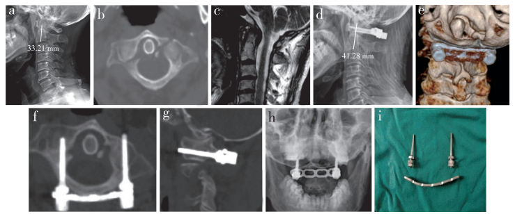
2 结果所有手术顺利完成。患者随访6 ~ 72个月,平均41个月,均获得骨性融合。手术时间为80 ~ 120 min,平均76 min;术中出血量为100 ~ 300 mL,平均150 mL。术后1周、末次随访时LMD、R-J线、VAS评分、JOA评分均较术前明显改善,差异有统计学意义(P < 0.05,表 1)。1例术前Frankel分级D级患者术后恢复至E级;其余24例仍为E级。术中2例患者在剥离寰椎后弓下缘时损伤静脉丛,用明胶海绵压迫止血;术后1例患者CT检查示螺钉突破椎弓根内侧骨皮质,但无神经症状。所有患者末次随访时未见明显复位丢失,钢板内固定在位、牢靠。典型病例影像学资料见图 1。
|
|
表 1 评价指标 Tab. 1 Evaluation index |

|
男,54岁,寰椎后弓合并侧块骨折,术前JOA评分9分,Frankel分级D级 a:术前颈椎X线片示R-J线33.21 mm b:术前CT示寰椎前弓及对侧侧块骨折 c:术前MRI示颈脊髓轻度受压 d:术后X线片示R-J线恢复至41.28 mm e ~ g:术后CT示骨折对位对线良好、骨折愈合、内固定稳定 h:术后1年张口位X线片示内固定器位置良好 i:术中所用颈椎后路钉板系统 Male, 54 years old, posterior arch and lateral mass fracture, preoperative JOA score is 9, Frankel classification is D a:Preoperative cervical reontgenograph shows R-J line is 33.21 mm b:Preoperative CT shows anterior arch of atlas and contralateral lateral mass fracture c:Preoperative MRI shows light cord signals of spine d:Postoperative reontgenograph shows that R-J line recovers to 41.28 mm e-g:Postoperative CTs show good contralateral alignment, fracture healing and stable internal fixation h:Mouth-open roentgenograph at postoperative 1 year shows that internal fixation is in good position i:Intraoperative cervical posterior nailboard system 图 1 典型病例影像学资料 Fig. 1 Imaging data of a typical case |
不稳定性寰椎骨折危险系数较高,如果对脊髓造成卡压,将对患者生命健康及生活质量造成严重威胁。因此,选择适宜的治疗方式是提升患者生存质量的关键,而维持寰枢关节的稳定性、避免因不稳而导致的延髓受压是治疗的最终目的。生理性固定技术是可以兼顾恢复寰椎稳定性与保留活动功能的手术方法,其目前主要用于不伴有横韧带断裂的寰椎骨折。对于横韧带断裂患者,也可获得良好的临床效果[10]。经椎弓根螺钉内固定作为一种生理性固定手术,仅处理损伤的寰椎,不涉及毗邻骨骼及韧带,还能迅速使寰椎达到解剖复位,并有效维持颈椎稳定性,有利于解除脊髓及神经压迫,并防止进一步损伤,主要适用于不稳定但横韧带完整的寰椎骨折,同时也可以用于部分横韧带断裂的寰椎骨折,慎用于粉碎性侧块骨折、前弓多处骨折及典型的Jefferson骨折。本研究25例患者均采用经椎弓根螺钉固定,通过安装预弯钢板,加压内聚,使分离的侧块直接复位,所有寰椎均获得良好复位,术后颈椎序列良好,骨折断端获得骨性融合,枕-寰-枢复合体高度明显恢复,未见明显失稳现象,疼痛缓解,颈部活动范围基本接近伤前水平,临床疗效良好。目前越来越多的学者主张在条件许可的情况下尽可能减少固定融合节段[11],关注点开始从骨折本身向骨折愈合后稳定结构的功能转变。
从力学角度上,重建寰椎环的稳定性可以利用纵行韧带的牵拉恢复枕-寰-枢复合体的生理高度。R-J线是指枢椎椎体下缘中点到基底线的垂直距离,相关研究报道R-J线可作为上颈椎病变齿突垂直移位、颅骨沉降的指标[12],由此可推想,在寰椎骨折中,枢椎及颅骨均完好,可近似看作是颅骨与枢椎之间的距离变化,进而可利用R-J线的测量值来近似代表枕-寰-枢复合体的高度,以此为量化值来衡量骨折前后枕-寰-枢复合体高度的变化。寰椎骨折后寰椎环失稳,受力以向外分散力为主,枕-寰-枢复合体高度下降,垂直应力瞬间增强,C0~2高度下降,轴向的压力迫使寰椎向两侧分离移位,致使关节面外移,寰枕关节面失吻合,R-J线缩短,进而导致关节活动范围变小、产生疼痛。本研究组病例术后随访LMD改善率达68.41%,R-J线较术前恢复35.40%,疼痛程度及功能活动均较术前显著改善,间接反映了术中复位越好,骨折间隙较窄的患者术后R-J线的恢复程度相对较高,同时后期患者的上颈椎活动度相对较好,证明R-J线可以用来评价患者骨折的复位情况,也可以预测术后的功能活动恢复情况。不足之处在于R-J线仅在术前、术后侧位X线片上评价寰枢椎复合体高度,并没有进行数学模型重建、复原原高度而进一步评价,因此还需进一步研究来验证R-J线评价枕-寰-枢复合体高度的合理性。
综上,后路椎弓根螺钉单节段内固定治疗寰椎骨折,可重建寰椎稳定性,恢复枕-寰-枢复合体高度,同时也最大限度保留了颈椎活动度,提高患者术后生活质量。但单节段固定不是治疗寰椎骨折的唯一方法,临床中还需综合考虑患者的具体情况及医疗单位的条件合理选择手术方法,以期获得最优疗效。
| [1] |
Li L, Teng H, Pan J, et al. Direct posterior c1 lateral screws compression reduction and osteosynthesis in the treatment of unstable Jefferson fractures[J]. Spine(Phila Pa 1976), 2011, 36(15): E1046-E1051. DOI:10.1097/BRS.0b013e3181fef78c |
| [2] |
Tan J, Li L, Sun G, et al. C1 lateral mass-C2 pedicle screws and cross link compression fixation for unstable atlas fracture[J]. Spine(Phila Pa 1976), 2009, 34(23): 2505-2509. DOI:10.1097/BRS.0b013e3181b4009a |
| [3] |
王迎松, 刘路平, 张颖, 等. C1, 2椎弓根钉棒固定治疗寰椎骨折(Jefferson骨折)疗效分析[J]. 脊柱外科杂志, 2010, 8(1): 1-3. |
| [4] |
胡勇, 马维虎, 顾勇杰, 等. 经口咽入路内固定治疗孤立性寰椎骨折临床疗效分析[J]. 脊柱外科杂志, 2011, 9(3): 131-134. |
| [5] |
马向阳, 尹庆水, 吴增辉, 等. 寰椎椎弓根与枢椎侧块关系的解剖与临床研究[J]. 中华骨科杂志, 2004, 24(5): 42-45. |
| [6] |
Fukui M, Chiba K, Kawakami M, et al. Japanese Orthopaedic Association cervical myelopathy evaluation questionnaire(JOACMEQ):Part 2. Endorsement of the alternative item[J]. J Orthop Sci, 2007, 12(3): 241-248. DOI:10.1007/s00776-007-1119-0 |
| [7] |
Huskisson EC. Measurement of pain[J]. Lancet, 1974, 2(7889): 1127-1131. |
| [8] |
Frankel HL, Hancock DO, Hyslop G, et al. The value of postural reduction in the initial management of closed injuries of the spine with paraplegia and tetraplegia. I[J]. Paraplegia, 1969, 7(3): 179-192. |
| [9] |
韩岳, 马信龙, 夏群, 等. 颈椎类风湿性关节炎的临床特点及诊疗进展[J]. 中华骨科杂志, 2014, 34(9): 962-967. DOI:10.3760/cma.j.issn.0253-2352.2014.09.011 |
| [10] |
He B, Yan L, Zhao Q, et al. Self-designed posterior atlas polyaxial lateral mass screw-plate fixation for unstable atlas fracture[J]. Spine J, 2014, 14(12): 2892-2896. DOI:10.1016/j.spinee.2014.04.020 |
| [11] |
胡勇, 董伟鑫, 徐荣明. 上颈椎内固定技术的研究近况[J]. 脊柱外科杂志, 2016, 14(4): 252-256. |
| [12] |
Redlund-Johnell I, Pettersson H. Vertical dislocation of the C1 and C2 vertebrae in rheumatoid arthritis[J]. Acta Radiol Diagn(Stockh), 1984, 25(2): 133-141. |
 2019, Vol.17
  Issue(6): 379-382


