 2020, Vol.18
  Issue(2): 77-81


腰椎峡部裂的主要临床表现为腰痛,偶有下肢放射痛,在青少年中发生率较高,是青少年腰痛的常见原因,而疲劳断裂被认为是其发生的主要原因[1-4]。腰椎峡部裂的风险因素研究显示,50%峡部裂患者有不同程度的椎体滑脱,因此,峡部裂的早期治疗对预防和缓解椎体滑脱非常重要[5]。对经非手术治疗无效的腰椎峡部裂患者及时实施修复手术,可有效减轻疼痛、促进峡部裂愈合、防止椎体滑脱以及控制其发展。既往研究采用峡部清理后取髂骨植骨并跨运动节段内固定治疗腰椎峡部裂,愈合率高、并发症少、内固定取出后固定节段仍存在有效活动,临床疗效满意[6]。但在手术过程中发现,常规显露方法需要剥离附着在椎板上的多裂肌,使峡部周围血供进一步减少,峡部裂患者椎弓根进钉点往往更靠近外侧,需要更长的切口显露才能获得满意的置钉角度。2014年10月—2016年10月本院采用经Wiltse入路[7]完成峡部清理后取髂骨植骨并跨运动节段内固定治疗峡部裂患者48例,临床疗效满意,现报告如下。
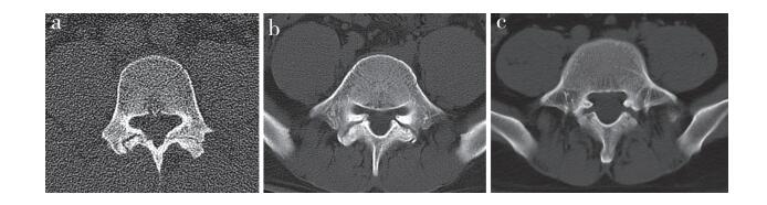
1 资料与方法 1.1 一般资料纳入标准:①矢状位CT三维重建确诊为腰椎峡部裂;②单节段腰椎峡部裂,若合并腰椎滑脱则Meyerding分级[8]应≤Ⅰ度;③症状以腰痛为主,疼痛视觉模拟量表(VAS)评分[9] > 5分,无持续下肢放射痛,无下肢神经受压症状;④非手术治疗 > 6个月症状无缓解,佩戴支具 > 6个月腰椎峡部裂未愈合;⑤经双侧峡部封闭(局部麻醉药物)治疗,腰痛缓解 > 50%;⑥固定节段椎间盘退行性变Pfirrmann分级[10]≤Ⅲ级;⑦无既往腰椎手术史。根据上述标准纳入患者48例,其中男42例、女6例,年龄17 ~ 35(22.0±3.5)岁。病变节段:L3 4例,L4 6例,L5 38例。临床表现均以腰痛为主要症状,腰部后伸、旋转后疼痛加重,休息或平卧可缓解。查体:单侧或双侧棘突旁、棘突推挤痛,关节突处压痛,腰部屈伸、旋转活动部分受限,双下肢感觉、运动、肌力、腱反射正常。所有患者入院后均行腰椎正侧位、前屈后伸位、双斜位X线,CT三维重建及MRI检查。根据术前腰椎峡部裂CT表现分为3型[11](图 1):Ⅰ型(线型)15例,峡部裂缝隙狭窄,类似于线性,更倾向于峡部骨折;Ⅱ型(中间型)24例,峡部裂缝隙较大,存在一定缺损,但未见假关节及骨硬化表现;Ⅲ型(硬化型)9例,峡部裂间隙明显,断端可见硬化,峡部裂呈假关节表现。
|
a:Ⅰ型b:Ⅱ型c:Ⅲ型 a:Type Ⅰ b:Type Ⅱ c:Type Ⅲ 图 1 腰椎峡部裂CT分型 Fig. 1 CT classification of lumbar spondylolysis |
患者全身麻醉后取俯卧位,透视确认腰椎后正中切口体表标记,切开皮肤、皮下组织,双侧棘突旁约2 cm处纵行切开腰背筋膜(切口长度约7 cm),显露椎旁竖脊肌群,寻找多裂肌与最长肌的肌间隙,经Wiltse入路显露进钉点,于峡部裂头侧及尾侧椎弓根内置入合适的椎弓根螺钉。仔细剥离峡部表面的多裂肌,显露峡部裂区域,将峡部的纤维瘢痕组织彻底刮除,磨钻磨除峡部断端硬化骨直至骨断面粗糙渗血。同一切口内,在髂后上棘开窗后用刮匙刮除松质骨,填塞于清理完毕的峡部裂隙中,并用植骨棒打压结实,剩余自体骨覆盖在峡部及横突周围。选择合适长度的连接杆,适当预弯后与椎弓根螺钉连接并锁紧,检查植骨处稳定后关闭切口。术后1 d拔除负压引流管,术后2 d佩戴腰部支具下床活动,活动量循序渐进,腰部支具佩戴3个月。
1.3 评价指标术后定期行腰椎X线及CT三维重建检查,影像学资料显示峡部间隙消失、出现连续骨痂为判断愈合的标准,并记录愈合时间。双侧峡部愈合后取出内固定,取出内固定的患者行腰椎前屈后伸位X线检查,评估固定节段的活动情况。采用VAS评分评估腰部及髂骨取骨部位疼痛程度,采用日本骨科学会(JOA)评分[12]评估腰椎功能。
1.4 统计学处理采用SPSS 15.0软件对数据进行统计学分析,计量资料以x±s表示,采用配对样本t检验;以P < 0.05为差异有统计学意义。
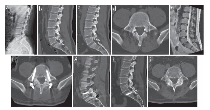
2 结果所有手术顺利完成。手术时间90 ~ 180 min,平均107 min;术中出血量50 ~ 160 mL,平均90 mL。所有患者随访6 ~ 36(15.3±3.8)个月,48例患者共96侧峡部裂,术后3个月愈合9侧,其中双侧愈合2例,单侧愈合5例;术后6个月愈合22侧,其中双侧愈合7例,单侧愈合8例;术后9个月愈合56侧,其中双侧愈合22例,单侧愈合12例;术后1年愈合83侧,其中双侧愈合38例,单侧愈合7例;术后2年愈合91侧,其中双侧愈合45例,单侧愈合1例。术后1年愈合率为86.5%(83/96),术后2年愈合率为94.8%(91/96);5侧未愈合,双侧未愈合2例,单侧未愈合1例,均为Ⅲ型患者。腰痛VAS评分由术前(6.9±2.7)分降至末次随访时(1.5±0.3)分,取髂骨部位疼痛VAS评分由术后1个月(3.7±1.3)分降至末次随访时(0.9±0.4)分;腰椎JOA评分由术前(20.3±3.2)分恢复到末次随访时(26.9±1.0)分,差异均有统计学意义(P < 0.05)。术后1年,22例患者取出内固定,取出后运动节段活动度为4° ~ 10°(5.6°±2.3°)。术后切口延迟愈合1例,其余无脑脊液漏、神经损伤、感染等其他并发症发生。典型病例影像学资料见图 2。
|
男,34岁,腰痛2年a ~ d:术前X线片和CT示双侧L5峡部裂,CT分型Ⅲ型,L5Ⅰ度滑脱e:术前腰椎MRI示L5/S1椎间盘退行性变Pfirrmann分级Ⅱ级f:术后即刻CT示峡部间隙变小,植骨块位置良好g、h:术后18个月CT示峡部骨性愈合i:内固定取出后CT示峡部愈合牢固 Male, 34 years old, back pain for 2 years a-d:Preoperative roentgenograph and CTs show bilateral L5 isthmus, CT classification typeⅢ, L5Ⅰdegree spondylolisthesis e:Preoperative MRI shows L5/S1 disc degeneration Pfirrmann GradeⅡ f:CT immediately after operation shows defect space became smaller and bone graft is in good position g, h:Postoperative 18 months CTs show isthmus bone healing i:After removal of internal fixation, CT shows firm healing of isthmus 图 2 典型病例影像学资料 Fig. 2 Imaging data of a typical case |
腰痛是腰椎峡部裂的主要症状,非手术治疗是主要治疗方法,大多数患者疗效满意[13]。手术治疗是在非手术治疗无效时的选择。本研究中患者腰痛均持续 > 6个月,且经腰围保护6个月无效后选择手术治疗。有研究报道,70%双侧峡部裂患者伴有不同程度的椎体滑脱,部分滑脱需要手术治疗,因此,峡部裂的早期治疗对预防椎体滑脱非常重要[14]。因早期缺乏内固定手段,峡部裂最早的手术治疗仅仅是峡部植骨原位融合后长期卧床等待愈合。1994年,胥少汀[15]报道使用经峡部螺钉内固定治疗峡部裂,最大的问题是螺钉通过峡部减少了植骨面积,且螺钉断裂发生率较高。2005年,李放等[1]报道使用椎弓根螺钉-椎板钩内固定治疗单纯腰椎峡部裂,固定范围局限在单一椎体,克服了融合手术的弊端,但个别患者L5椎板窄薄,椎板钩放置不合适,只能选择其他单椎体内固定方式,如横突棘突钢丝捆绑内固定术(Scott法)、椎弓根螺钉-“U”形棒法等[16],均获得满意疗效,但抗旋转、后伸能力较弱。本研究组采用峡部清理取髂骨植骨跨运动节段内固定治疗腰椎峡部裂,融合率高,并发症发生率低,内固定取出后仍能保留固定节段部分活动度,证实跨运动节段内固定能提供更强的稳定性,也提示该法是一种可靠的治疗方法[6]。
常规后正中入路需要剥离双侧骶棘肌,通常使用电刀将多裂肌自椎板完全剥离,肌肉损伤大,易导致肌肉的失神经支配,破坏腰椎后方肌肉韧带稳定结构[17]。虽然椎板及峡部剥离较为光整,但峡部裂局部背侧的软组织完全剥离致使背侧血运完全被破坏,仅残留腹侧血运,不利于峡部裂愈合[18]。为减少对峡部血运的破坏,本研究组经后正中入路切开皮肤、皮下组织,然后自两侧切开腰背筋膜,寻找多裂肌与最长肌的肌间隙,经Wiltse入路仅显露峡部裂区域,尽可能保护峡部的血运,以提高峡部愈合率。既往手术中发现,峡部裂患者的椎弓根进钉点更偏向外侧,被峡部增生的骨赘所遮挡,往往需要更多头尾侧软组织的剥离显露才能找到满意的进钉点和角度。Wiltse入路经最短距离直达椎弓根螺钉的进钉点,减少了显露的长度、时间及术中出血量,可提供更佳的置入角度和长度,固定更加牢靠。而且,通过Wiltse入路置钉,螺钉尾部可以远离峡部植骨区域,从而减少对局部的干扰。本研究组患者术中出血量较小,切口长度比常规显露切口短约2 cm,患者术后的切口疼痛和不适也较轻,总体上减小了手术创伤。与本研究组既往研究[6]比较发现,Wiltse入路术后各时间点峡部愈合率略高于传统入路,但差异无统计学意义;术中出血量小于传统入路,差异具有统计学意义。
另外,峡部裂修复手术的关键步骤是峡部瘢痕的清理,可使用磨钻磨除硬化的峡部断端,直至出现粗糙渗血的植骨床。手术需要取髂骨,常规传统入路通常是在切口内切开髂嵴后方的筋膜取骨,经Wiltse入路可在肌间隙入路内找到髂嵴而直接完成取骨,不需要额外显露,减少了损伤。该入路的不足之处:向内侧剥离多裂肌,峡部裂最内侧区域往往显露不足,造成内侧峡部的清理不足,但从本研究的术后愈合情况来看,并没有产生太大的影响。
综上,经Wiltse入路峡部植骨跨运动节段内固定治疗腰椎峡部裂,术中出血量小、术后引流量少、患者恢复快、切口疼痛和不适较轻、峡部愈合率高、并发症发生率低,在修复峡部裂的同时可保留峡部血运,值得应用推广。
| [1] |
李放, 关凯, 戴刚, 等. 椎弓根钉-椎板钩系统固定治疗单纯腰椎峡部裂[J]. 中国脊柱脊髓杂志, 2005, 15(11): 9-11. |
| [2] |
Wiltse LL, Widell EH Jr, Jackson DW. Fatigue fracture:the basic lesion is inthmic spondylolisthesis[J]. J Bone Joint Surg Am, 1975, 57(1): 17-22. DOI:10.2106/00004623-197557010-00003 |
| [3] |
Sairyo K, Katoh S, Sasa T, et al. Athletes with unilateral spondylolysis are at risk of stress fracture at the contralateral pedicle and pars interarticularis:a clinical and biomechanical study[J]. Am J Sports Med, 2005, 33(4): 583-590. DOI:10.1177/0363546504269035 |
| [4] |
林云飞, 王飞, 李放. 导致腰椎峡部裂的风险因素研究现状[J]. 脊柱外科杂志, 2016, 14(3): 181-184. DOI:10.3969/j.issn.1672-2957.2016.03.011 |
| [5] |
Wiltse LL, Newman PH, Macnab I. Classification of spondylolisis and spondylolisthesis[J]. Clin Orthop Relat Res, 1976(117): 23-29. |
| [6] |
张志成, 李放, 孙天胜, 等. 峡部植骨修复运动节段固定治疗青少年腰椎峡部裂研究[J]. 中华骨与关节外科杂志, 2018, 11(9): 651-655. DOI:10.3969/j.issn.2095-9958.2018.09.003 |
| [7] |
Vialle R, Wicart P, Drain O, et al. The Wiltse paraspinal approach to the lumbar spine revisited:an anatomic study[J]. Clin Orthop Relat Res, 2006, 445: 175-180. |
| [8] |
Meyerding HW. Low backache and sciatic pain associated with spondylolisthesis and protruded intervertebral disc:incidence, significance and treatment[J]. J Bone Joint Surg Am, 1941, 23(2): 461-470. |
| [9] |
Huskisson EC. Measurement of pain[J]. Lancet, 1974, 2(7889): 1127-1131. |
| [10] |
Castro-Mateos I, Hua R, Pozo JM, et al. Intervertebral disc classification by its degree of degeneration from T2-weighted magnetic resonance images[J]. Eur Spine J, 2016, 25(9): 2721-2727. DOI:10.1007/s00586-016-4654-6 |
| [11] |
Sairyo K, Sakai T, Yasui N, et al. Conservative treatment for pediatric lumbar spondylolysis to achieve bone healing using a hard brace:what type and how long? Clinical article[J]. J Neurosurg Spine, 2012, 16(6): 610-614. DOI:10.3171/2012.2.SPINE10914 |
| [12] |
Fukui M, Chiba K, Kawakami M, et al. Japanese Orthopaedic Association Back Pain Evaluation Questionnaire. Part 2. Verification of its reliability:the Subcommittee on Low Back Pain and Cervical Myelopathy Evaluation of the Clinical Outcome Committee of the Japanese Orthopaedic Association[J]. J Orthop Sci, 2007, 12(6): 526-532. DOI:10.1007/s00776-007-1168-4 |
| [13] |
Sakai T, Tezuka F, Yamashita K, et al. Conservative treatment for bony healing in pdiatric lumbar spondylolysis[J]. Spine(Phlia Pa 1976), 2017, 42(12): E716-E720. DOI:10.1097/BRS.0000000000001931 |
| [14] |
Sakai T, Sairyo K, Takao S, et al. Incidence of lumbar spondylolysis in the general population in Japan based on multidetector computed tomography scans from two thousand subjects[J]. Spine(Phlia Pa 1976), 2009, 34(21): 2346-2350. DOI:10.1097/BRS.0b013e3181b4abbe |
| [15] |
胥少汀. 腰椎峡部裂与脊椎滑脱[J]. 中国矫形外科杂志, 1994, 1(2): 103-105. |
| [16] |
朱磊, 许鹏, 王冬冬, 等. 青少年腰椎峡部裂单椎体内固定研究进展[J]. 脊柱外科杂志, 2019, 17(3): 211-215. DOI:10.3969/j.issn.1672-2957.2019.03.013 |
| [17] |
Boelderl A, Daniaux H, Kathrein A, et al. Danger of damaging the medial branches of the posterior rami of spinal nerves during a dorsomedian approach to the spine[J]. Clin Anat, 2002, 15(2): 77-81. |
| [18] |
Vaccaro AR, Chiba K, Heller JG, et al. Bone grafting alternatives in spinal surgery[J]. Spine J, 2002, 2(3): 206-215. DOI:10.1016/S1529-9430(02)00180-8 |